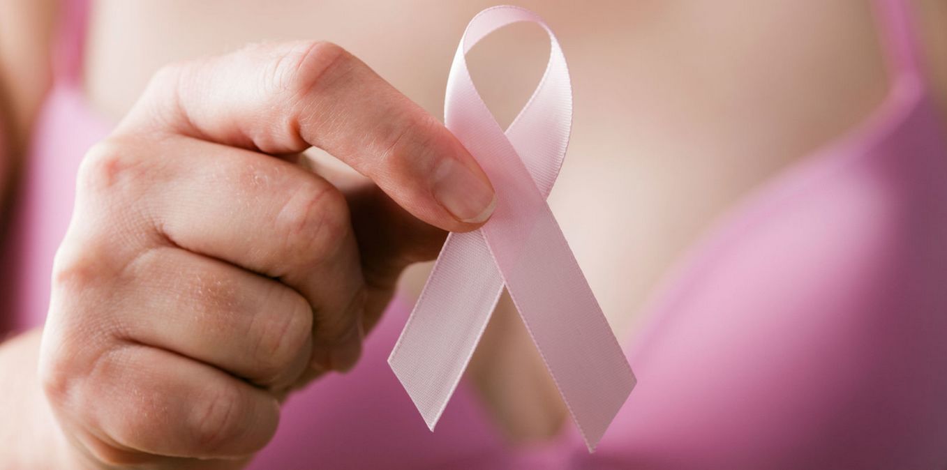
Second Life is in the midst of the 2018 Relay for Life season, most notably (at the time of writing) with Fantasy Faire. Given that it is, I would like to step to one side from my usual writing and offer a personal piece on the subject of cancer. It’s something I’ve spent a couple of days wrestling over committing to print, and I’m now doing so not to illicit sympathy, but to hopefully offer insight into why it’s better to confront things then shy away from things out of fear of hearing the “c” word.
Earlier this year I was diagnosed with DCIS – ductal carcinoma in situ – in my left breast. This is a form of best cancer where the cancerous cells are contained within the milk ducts of the breast. Because the cancer cells have not invaded nearby breast tissue, DCIS is regarded as non-invasive breast cancer, and accounts for about 20% of all breast cancer cases, and around 85% of all in-situ (confined to a specific area) forms of breast cancer.
While there is a risk it might become invasive if left untreated (the American Cancer Society estimate between 20-53% of untreated in-situ cancer cases become invasive over a period of about a decade, DCIS can be dealt with in a relatively straightforward manner through what amounts to a two-step treatment process.
The first step is for the affected area of breast duct to be surgically removed in a localised procedure referred to as a lumpectomy. This is a form of surgery designed to excise the affected area, and as a rule leaves the breast looking as close as possible to how it did before surgery, with its general shape and the nipple area remaining intact.
After a time for healing, the second step is generally a period of localised radiotherapy. This is designed to destroy any remaining cancer cells that would otherwise by too small to see on scans or to measure with lab tests. In addition, it can lower both the risk of DCIS returning to the breast, or of the breast developing an invasive cancer later in life.

Obviously, “surgery” and “radiotherapy” are themselves terrifying words; but the fact is that often, DCIS can be dealt with on an out-patient basis – there’s no need for a protracted stay in hospital; while the radiotherapy is localised enough such that the risk of it giving rise to cancer later in life is around 5% – far less a risk than that of the DCIS leading to a more invasive form of cancer.
A key point with DCIS is that it is hard to detect; while it may be indicated by a subcutaneous lump, often it is only through a scan and / or biopsy that it may be identified. In my case, I noticed a small lump in my right breast; when it hadn’t gone away after a number of weeks, I went to see my GP.
I admit, my feelings were mixed when I did so: cancer has been a frequent visitor within both sides of my family, so I was concerned I would hear the words “breast cancer”; at the same time, there was also a feeling that I was “just being silly” and over-reacting to something that would go away – after all, lumps in the breast can be caused by a lot of non-cancerous events.

In fact, the right breast lump did prove to be a small non-cancerous node of breast calcification. However, as a result of the scans my GP sent me to have, the left breast DCIS was spotted.
Cutting a long story short, I was referred for surgery at the cancer unit of a local hospital, where I underwent two bouts of surgery some 14 days apart. The first was to excise the affected ductal area, the second to remove a small amount of tissue from the surrounding area. Both bouts of surgery were performed on an out-patient basis, so I went into hospital in the morning and was back home and in my own bed in the evening.
After the surgery I had several weeks of recovery to allow the surgical wound and the (admittedly extensive) bruising around it to heal. I have been left with a scar marking the entry wound, but the shape of my breast hasn’t changed and as is common with this type of surgery, the scar itself is on the underside of the breast, so it’s not naturally visible.
As to the radiotherapy, I was given 15 sessions broken down over just over three workday weeks, plus an initial “targeting” session a week ahead of the treatment. The treatment took the form of spirometry-monitored deep inspiration breath hold (SMDIBH). Again this sounds a mouthful, and possibly frightening, but what it amounts to is being subjected to a short burst of radiation while controlling you breathing and holding your breath for around 20-30 seconds. This approach is used when treating left breast cancer, as filling the lungs with air raises the breast away from the heart, reducing the amount of radiation to which the heart is exposed.
The treatment itself is quite painless, each “zap” lasting around 20 seconds as the breath is held, with the number of zaps you get varying according to need. However, due to the frequency of the treatment sessions, there are side-effects. These can include fatigue; a swelling in the breast due to fluid being unable to drain properly; a reddening and drying of the skin around the treated area, and a gradual feeling of heat build-up in the breast which takes time to dissipate. I found these symptoms took several weeks to abate, and was recommended to use a non-metallic moisturising cream to ease the dryness / discomfort.
As I write this, I’m into my second week of post-radiotherapy recovery. I’ll make no bones about it, my breast is sore I’m at times in a little discomfort and have felt lethargic at times – an effect that should subside over the next few weeks. However, the preliminary results of the treatment is that the surgery has been successful, and the radiotherapy will have hopefully done its job.
So why tell you all this? Because – as I said at the top, cancer’s biggest weapon is fear – fear of what it might mean if diagnosed and, equally, the fear of learning you have it in the first place. Yet the fact is, as my case hopefully shows, getting diagnosed early enough not only means a better chance of dealing with it – it also means the treatment is often less protracted and invasive than might otherwise be the case (put it this way, while it may well sound worrying when first heard, a lumpectomy is, overall, a lot less traumatic than a mastectomy) – whereas the longer it is ignored in the hope it might “go away” or because it spares us having to confront it, the greater the risk that it might reach a point were it cannot be more effectively dealt with.
Cancer is not something we can avoid simply by ignoring the signs (when they are present) or by avoiding the opportunity to have it diagnosed. So please, if you have concerns about anything, a lump here or there, a mole-like mark on your skin that has appeared or which has changed in size or has been subject to bleeding – go and get it checked. It might be cancer – or it might be something else entirely; it might be entirely benign. But if you don’t get it checked, you run the risk of not knowing – or of receiving medical help at a time when, should it prove to be cancer, it might be more easily dealt with than might be the case if you just ignore it.
In my case, I’m grateful I didn’t let the feeling of “being silly” when going to see my GP get the better of me; as a woman in my 40’s (no, I’m not saying where in my 40s!) I’m still several years from my first routine breast cancer screenings, possibly time enough for the DCIS to have become more of a problem. As it is, it’s now excised, and I’ll be having regular scans to make sure it stays that way. And that’s a form of peace of mind I’m grateful to have.
So again, if you have a suspicion or concern, don’t leave it for “another day”; go get it seen to.
